

Diagnostic Imaging · Dental Radiography
Portable Veterinary Dental X-ray
A lightweight, high-frequency handheld dental X-ray unit paired with a digital sensor — chairside intraoral radiography for the two-thirds of dental disease that lives below the gum line.
Why portable dental X-ray
Dental radiography changes what you find and what you treat — and a handheld unit brings it to any table, chair or exotic patient without a dedicated imaging room.
Truly portable
At roughly 2.2 kg with battery power and a universal AC input, it moves to the patient — general table, dental suite, or an exotic case — no fixed installation.
Sharp, fast exposures
A 0.4 × 0.4 mm focal spot and 60 kV / 2.0 mA high-frequency output with 0.1–1.2 s exposures capture crisp intraoral detail — roots, bone and periodontal structures.
Digital workflow
Paired with a Pluto digital sensor and imaging software, exposures become instant on-screen images ready to review, annotate and store in the patient record.
How it works
A simple chairside intraoral workflow — from positioning to a digital image in seconds.
- Position the unit. Aim the handheld tube head at the target tooth; the 130 mm focus-to-skin distance and light frame make positioning quick.
- Place the sensor. Position the digital sensor intraorally behind the target tooth or region.
- Expose. Trigger a short 0.1–1.2 s high-frequency exposure at 60 kV.
- Review instantly. The digital sensor renders the image on-screen for immediate assessment and annotation.
- Store to the record. Save images to the patient’s dental chart in NXVET (see below).
See it in action
Watch the handheld dental X-ray in a real veterinary dental workflow.
Product demo video
Coming soon
Where clinics use it
COHAT & dental cleaning
Full-mouth radiographs during comprehensive oral health assessment reveal disease hidden below the gum line.
Extraction planning
Image root anatomy before extractions to plan the approach and confirm complete removal.
Feline resorptive lesions
Detect and stage tooth resorption that isn’t visible on oral exam alone.
Endodontics & pathology
Assess root canals, periapical lucencies, fractures and bone loss.
Exotic & small patients
Portability suits rabbits, rodents and exotics where a fixed unit can’t reach.
Referral & mobile practice
Battery operation supports house-call and mobile dentistry.
Specifications
Handheld high-frequency dental X-ray unit (PETFECT TT-32DX), designed for pets and paired with a digital sensor.
| Tube voltage | 60 kV | Tube current | 2.0 mA |
| Exposure time | 0.1–1.2 s | Frequency | 30 kHz (high-frequency DC) |
| Tube focal spot | 0.4 × 0.4 mm | Focus-to-skin | 130 mm |
| Rated power | 120 VA | Battery | 2200 mAh |
| Input voltage | AC 100–240 V ±10% | Net weight | 2.2 kg |
| Dimensions | 170 × 153 × 138 mm | Operating temp. | 5–40 °C |
Pairs with a Pluto digital intraoral sensor and imaging software (models 0003X / 0004X). Specifications are indicative and confirmed per manufacturer at time of order.
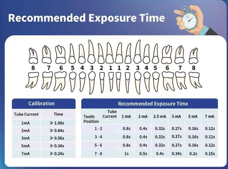
Recommended exposure guide
Suggested exposure times by tooth position and tube current — a practical chairside reference for consistent diagnostic images.
Handheld vs. fixed / wall-mount X-ray
| Portable handheld | Fixed / wall-mount | |
|---|---|---|
| Installation | None — use anywhere | Dedicated room & mounting |
| Mobility | Moves to the patient & house-calls | Patient must come to the unit |
| Exotic / small patients | Easy access & angles | Harder to position |
| Upfront footprint | Minimal | Higher build-out |
Handheld units are used following each manufacturer’s radiation-safety guidance and local regulations.
Works with the NXVET cloud
Dental images on the chart — not stuck on a workstation.
- Store intraoral radiographs directly on the patient’s dental record in NXVET.
- Compare images across visits to track periodontal and root pathology over time.
- Share radiographs with owners and referral partners from one record.
- Build a connected imaging + diagnostics stack alongside your other NX-supplied equipment.
Frequently asked questions
Is it truly portable?
What image quality does it produce?
Do I need a separate sensor?
What about radiation safety?
Can images go into our records?
Related products
Bring dental imaging chairside
Request a quote or the full TT-32DX and sensor specification, and we’ll help you set up a digital dental radiography workflow.